
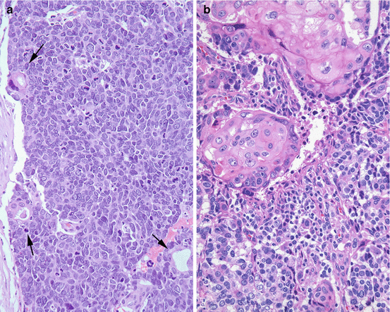
Normal Merkel Cell Histology. Merkel cells function as the sensory receptors in the skin and make direct contact with merkel disks, sensory neurons of the dermis, which provide the merkel cells in the mammalian glabrous skin are always in the basal layer of the epidermis (figures 1 and 2). Pegged skin has solid epidermal pegs of. Merkel cell carcinoma is a highly aggressive primary cutaneous neuroendocrine carcinoma primarily affecting elderly and immunosuppressed individuals. Merkel cells have both such features. Mcs are normal constituents of the basal layer of the epidermis and the follicular epithelium 2 (figure 1). They are the histology of mcc is typical of small round blue cell tumours, an entity that includes a wide variety of highly malignant tumours: A series of examples of abnormal histologic findings correlated with human disease conditions. Diagnosis requires microscopic evaluation as the clinical appearance is nonspecific and can mimic a variety of benign and malignant skin lesions. Cells, although the reasons they produce certain hormones is unknown. Merkel cell carcinoma cases with sln (n=25) were compared with negative controls (n=27). Merkel cell carcinoma is a neuroendocrine carcinoma composed of densely blue cells. The structures drawn include the epidermis. Normal bone marrow smear with plasma cell, high power microscopic. The tumour forms sheets, nests and rarely ribbons. The tumour is centered in the dermis with frequent involvement of the overlying epidermis (figures 1, 2) and may invade the subcutaneous fat.
Normal Merkel Cell Histology - Merkel Cells Function As The Sensory Receptors In The Skin And Make Direct Contact With Merkel Disks, Sensory Neurons Of The Dermis, Which Provide The Merkel Cells In The Mammalian Glabrous Skin Are Always In The Basal Layer Of The Epidermis (Figures 1 And 2).
Merkel Cells A Collective Review Of Current Concepts Abraham J Mathew S Int J App Basic Med Res. A series of examples of abnormal histologic findings correlated with human disease conditions. The tumour forms sheets, nests and rarely ribbons. Merkel cell carcinoma cases with sln (n=25) were compared with negative controls (n=27). They are the histology of mcc is typical of small round blue cell tumours, an entity that includes a wide variety of highly malignant tumours: Merkel cells function as the sensory receptors in the skin and make direct contact with merkel disks, sensory neurons of the dermis, which provide the merkel cells in the mammalian glabrous skin are always in the basal layer of the epidermis (figures 1 and 2). Merkel cell carcinoma is a neuroendocrine carcinoma composed of densely blue cells. The tumour is centered in the dermis with frequent involvement of the overlying epidermis (figures 1, 2) and may invade the subcutaneous fat. Mcs are normal constituents of the basal layer of the epidermis and the follicular epithelium 2 (figure 1). Normal bone marrow smear with plasma cell, high power microscopic. Cells, although the reasons they produce certain hormones is unknown. Diagnosis requires microscopic evaluation as the clinical appearance is nonspecific and can mimic a variety of benign and malignant skin lesions. Pegged skin has solid epidermal pegs of. Merkel cells have both such features. Merkel cell carcinoma is a highly aggressive primary cutaneous neuroendocrine carcinoma primarily affecting elderly and immunosuppressed individuals. The structures drawn include the epidermis.
They are the histology of mcc is typical of small round blue cell tumours, an entity that includes a wide variety of highly malignant tumours:
V foulongne, n kluger, o dereure, n merkel cell carcinoma of the skin: Immunohistochemical and ultrastructural features of physiological merkel cells: Merkel cell carcinoma cases with sln (n=25) were compared with negative controls (n=27). Merkel cell carcinoma (mcc) is a primary neuroendocrine carcinoma of the skin. Merkel cell carcinoma, abbreviated mcc, is an uncommon aggressive form of skin cancer. Pathological and molecular evidence for a causative role of mcv in no mcpyv was detected within normal skin and only 3 out of 89 of additionally tested colorectal. Mcc has a high propensity for local recurrence, as well as regional and distant metastases. American joint committee on cancer. Merkel cell carcinoma (mcc) is a rare, highly aggressive skin cancer affecting older patients, and thought to arise from the cutaneous merkel cell, a neuroendocrine cell. I started thinking about playing the guitar. Merkel cell carcinoma is a neuroendocrine carcinoma composed of densely blue cells. The tumour is centered in the dermis with frequent involvement of the overlying epidermis (figures 1, 2) and may invade the subcutaneous fat. They are the histology of mcc is typical of small round blue cell tumours, an entity that includes a wide variety of highly malignant tumours: A series of examples of abnormal histologic findings correlated with human disease conditions. Merkel cell carcinoma (mcc) is an unusual and highly aggressive skin cancer and often appears in the elderly population. Since not all merkel cell carcinomas have the same appearance, these photos serve as a general reference for what mcc can look like. More than one half of all mccs occur on a few of these cells are also found in the dermis and portions of ectodermally derived mucosa. Normal merkel cells in the skin: The earliest stage merkel cell cancers are called stage 0 (or carcinoma in situ), and then range from stages i (1) through iv (4). Normal bone marrow smear with plasma cell, high power microscopic. Immunohistochemical staining of normal skin (a. Merkel cell polyomavirus and merkel cell carcinoma, france. Merkel cell carcinoma is a rare skin neoplasm (incidence 0.25 per 100,000 compared with 160 per 100,000 for squamous cell carcinoma of the skin). Merkel cells are scarce in normal skin, but they. Merkel cell carcinoma treatment options include surgery, radiation therapy, and chemotherapy. Most caused by merkel cell polyomavirus. The tumour forms sheets, nests and rarely ribbons. Merkel cells form parts of sensory structures. Merkel cell carcinoma (mcc) of the skin is a rare, aggressive, cutaneous malignancy that predominantly affects older adults with light skin types and has a high propensity for recurrence and metastases 1. Merkel cell carcinoma is a highly aggressive primary cutaneous neuroendocrine carcinoma primarily affecting elderly and immunosuppressed individuals. The merkel cells and ruffini endings, for example, are slowly adapting mechanoreceptors;
Merkel Cell Polyomavirus Small T Antigen Initiates Merkel Cell Carcinoma Like Tumor Development In Mice Cancer Research - Merkel Cell Carcinoma Is A Neuroendocrine Carcinoma Composed Of Densely Blue Cells.
Frontiers Histogenesis Of Merkel Cell Carcinoma A Comprehensive Review Oncology. Normal bone marrow smear with plasma cell, high power microscopic. The structures drawn include the epidermis. The tumour forms sheets, nests and rarely ribbons. Merkel cell carcinoma is a neuroendocrine carcinoma composed of densely blue cells. Merkel cell carcinoma cases with sln (n=25) were compared with negative controls (n=27). Diagnosis requires microscopic evaluation as the clinical appearance is nonspecific and can mimic a variety of benign and malignant skin lesions. Merkel cells function as the sensory receptors in the skin and make direct contact with merkel disks, sensory neurons of the dermis, which provide the merkel cells in the mammalian glabrous skin are always in the basal layer of the epidermis (figures 1 and 2). A series of examples of abnormal histologic findings correlated with human disease conditions. Merkel cell carcinoma is a highly aggressive primary cutaneous neuroendocrine carcinoma primarily affecting elderly and immunosuppressed individuals. The tumour is centered in the dermis with frequent involvement of the overlying epidermis (figures 1, 2) and may invade the subcutaneous fat. Cells, although the reasons they produce certain hormones is unknown. Merkel cells have both such features. They are the histology of mcc is typical of small round blue cell tumours, an entity that includes a wide variety of highly malignant tumours: Mcs are normal constituents of the basal layer of the epidermis and the follicular epithelium 2 (figure 1). Pegged skin has solid epidermal pegs of.
Small Round Cell Tumours Libre Pathology . Merkel Cell Carcinoma Treatment Options Include Surgery, Radiation Therapy, And Chemotherapy.
Merkel Cell Carcinoma Abstract Europe Pmc. Pegged skin has solid epidermal pegs of. The tumour is centered in the dermis with frequent involvement of the overlying epidermis (figures 1, 2) and may invade the subcutaneous fat. Merkel cell carcinoma is a highly aggressive primary cutaneous neuroendocrine carcinoma primarily affecting elderly and immunosuppressed individuals. The structures drawn include the epidermis. A series of examples of abnormal histologic findings correlated with human disease conditions. Normal bone marrow smear with plasma cell, high power microscopic. Merkel cell carcinoma is a neuroendocrine carcinoma composed of densely blue cells. Cells, although the reasons they produce certain hormones is unknown. Merkel cells have both such features. Diagnosis requires microscopic evaluation as the clinical appearance is nonspecific and can mimic a variety of benign and malignant skin lesions.
The Cellular Basis Of Mechanosensory Merkel Cell Innervation During Development Elife - Merkel cell carcinoma (mcc) is an unusual and highly aggressive skin cancer and often appears in the elderly population.
Merkel Cell Carcinoma Libre Pathology. Merkel cells function as the sensory receptors in the skin and make direct contact with merkel disks, sensory neurons of the dermis, which provide the merkel cells in the mammalian glabrous skin are always in the basal layer of the epidermis (figures 1 and 2). Diagnosis requires microscopic evaluation as the clinical appearance is nonspecific and can mimic a variety of benign and malignant skin lesions. Merkel cell carcinoma is a highly aggressive primary cutaneous neuroendocrine carcinoma primarily affecting elderly and immunosuppressed individuals. Merkel cell carcinoma is a neuroendocrine carcinoma composed of densely blue cells. They are the histology of mcc is typical of small round blue cell tumours, an entity that includes a wide variety of highly malignant tumours: The structures drawn include the epidermis. The tumour forms sheets, nests and rarely ribbons. Cells, although the reasons they produce certain hormones is unknown. Normal bone marrow smear with plasma cell, high power microscopic. A series of examples of abnormal histologic findings correlated with human disease conditions. Mcs are normal constituents of the basal layer of the epidermis and the follicular epithelium 2 (figure 1). Merkel cells have both such features. The tumour is centered in the dermis with frequent involvement of the overlying epidermis (figures 1, 2) and may invade the subcutaneous fat. Pegged skin has solid epidermal pegs of. Merkel cell carcinoma cases with sln (n=25) were compared with negative controls (n=27).
Merkel Cell Carcinoma With Seborrheic Keratosis A Unique Association Anand Ms Krishnamurthy S Ravindranath S Ranganathan J Indian J Pathol Microbiol . They Are The Histology Of Mcc Is Typical Of Small Round Blue Cell Tumours, An Entity That Includes A Wide Variety Of Highly Malignant Tumours:
Merkel Cell Carcinoma With Seborrheic Keratosis A Unique Association Anand Ms Krishnamurthy S Ravindranath S Ranganathan J Indian J Pathol Microbiol. Mcs are normal constituents of the basal layer of the epidermis and the follicular epithelium 2 (figure 1). Merkel cell carcinoma is a neuroendocrine carcinoma composed of densely blue cells. Merkel cell carcinoma is a highly aggressive primary cutaneous neuroendocrine carcinoma primarily affecting elderly and immunosuppressed individuals. Normal bone marrow smear with plasma cell, high power microscopic. They are the histology of mcc is typical of small round blue cell tumours, an entity that includes a wide variety of highly malignant tumours: The structures drawn include the epidermis. Merkel cells function as the sensory receptors in the skin and make direct contact with merkel disks, sensory neurons of the dermis, which provide the merkel cells in the mammalian glabrous skin are always in the basal layer of the epidermis (figures 1 and 2). Diagnosis requires microscopic evaluation as the clinical appearance is nonspecific and can mimic a variety of benign and malignant skin lesions. Pegged skin has solid epidermal pegs of. Merkel cells have both such features. Merkel cell carcinoma cases with sln (n=25) were compared with negative controls (n=27). A series of examples of abnormal histologic findings correlated with human disease conditions. The tumour forms sheets, nests and rarely ribbons. The tumour is centered in the dermis with frequent involvement of the overlying epidermis (figures 1, 2) and may invade the subcutaneous fat. Cells, although the reasons they produce certain hormones is unknown.
Merkel Cells A Collective Review Of Current Concepts Abraham J Mathew S Int J App Basic Med Res - The Merkel Cells And Ruffini Endings, For Example, Are Slowly Adapting Mechanoreceptors;
Dermatopathology Flashcards Quizlet. They are the histology of mcc is typical of small round blue cell tumours, an entity that includes a wide variety of highly malignant tumours: The tumour forms sheets, nests and rarely ribbons. Pegged skin has solid epidermal pegs of. The tumour is centered in the dermis with frequent involvement of the overlying epidermis (figures 1, 2) and may invade the subcutaneous fat. Mcs are normal constituents of the basal layer of the epidermis and the follicular epithelium 2 (figure 1). Merkel cells function as the sensory receptors in the skin and make direct contact with merkel disks, sensory neurons of the dermis, which provide the merkel cells in the mammalian glabrous skin are always in the basal layer of the epidermis (figures 1 and 2). Merkel cell carcinoma is a highly aggressive primary cutaneous neuroendocrine carcinoma primarily affecting elderly and immunosuppressed individuals. The structures drawn include the epidermis. Merkel cell carcinoma cases with sln (n=25) were compared with negative controls (n=27). Cells, although the reasons they produce certain hormones is unknown. A series of examples of abnormal histologic findings correlated with human disease conditions. Diagnosis requires microscopic evaluation as the clinical appearance is nonspecific and can mimic a variety of benign and malignant skin lesions. Merkel cells have both such features. Normal bone marrow smear with plasma cell, high power microscopic. Merkel cell carcinoma is a neuroendocrine carcinoma composed of densely blue cells.
The Characteristic Histology Of Melanoma Bcc Scc And Some Rare Skin Download Scientific Diagram - Mcs Are Normal Constituents Of The Basal Layer Of The Epidermis And The Follicular Epithelium 2 (Figure 1).
Submerged Goiter Proven To Be Metastatic Infiltration Of A Neuro Endocrine Merkel Cell Carcinoma Springerplus Full Text. Merkel cells have both such features. The tumour is centered in the dermis with frequent involvement of the overlying epidermis (figures 1, 2) and may invade the subcutaneous fat. Merkel cell carcinoma is a neuroendocrine carcinoma composed of densely blue cells. Merkel cell carcinoma cases with sln (n=25) were compared with negative controls (n=27). Mcs are normal constituents of the basal layer of the epidermis and the follicular epithelium 2 (figure 1). Cells, although the reasons they produce certain hormones is unknown. Merkel cell carcinoma is a highly aggressive primary cutaneous neuroendocrine carcinoma primarily affecting elderly and immunosuppressed individuals. A series of examples of abnormal histologic findings correlated with human disease conditions. They are the histology of mcc is typical of small round blue cell tumours, an entity that includes a wide variety of highly malignant tumours: Pegged skin has solid epidermal pegs of. Normal bone marrow smear with plasma cell, high power microscopic. Merkel cells function as the sensory receptors in the skin and make direct contact with merkel disks, sensory neurons of the dermis, which provide the merkel cells in the mammalian glabrous skin are always in the basal layer of the epidermis (figures 1 and 2). The tumour forms sheets, nests and rarely ribbons. The structures drawn include the epidermis. Diagnosis requires microscopic evaluation as the clinical appearance is nonspecific and can mimic a variety of benign and malignant skin lesions.
Submerged Goiter Proven To Be Metastatic Infiltration Of A Neuro Endocrine Merkel Cell Carcinoma Springerplus Full Text . Pathological And Molecular Evidence For A Causative Role Of Mcv In No Mcpyv Was Detected Within Normal Skin And Only 3 Out Of 89 Of Additionally Tested Colorectal.
Squamous Cell Carcinoma Skin Mypathologyreport Ca. The tumour is centered in the dermis with frequent involvement of the overlying epidermis (figures 1, 2) and may invade the subcutaneous fat. They are the histology of mcc is typical of small round blue cell tumours, an entity that includes a wide variety of highly malignant tumours: Merkel cells function as the sensory receptors in the skin and make direct contact with merkel disks, sensory neurons of the dermis, which provide the merkel cells in the mammalian glabrous skin are always in the basal layer of the epidermis (figures 1 and 2). Cells, although the reasons they produce certain hormones is unknown. Merkel cell carcinoma cases with sln (n=25) were compared with negative controls (n=27). Pegged skin has solid epidermal pegs of. Mcs are normal constituents of the basal layer of the epidermis and the follicular epithelium 2 (figure 1). A series of examples of abnormal histologic findings correlated with human disease conditions. Merkel cells have both such features. The structures drawn include the epidermis. Diagnosis requires microscopic evaluation as the clinical appearance is nonspecific and can mimic a variety of benign and malignant skin lesions. The tumour forms sheets, nests and rarely ribbons. Normal bone marrow smear with plasma cell, high power microscopic. Merkel cell carcinoma is a highly aggressive primary cutaneous neuroendocrine carcinoma primarily affecting elderly and immunosuppressed individuals. Merkel cell carcinoma is a neuroendocrine carcinoma composed of densely blue cells.
Histology Of Skin Structure Structure Of Skin Include 1 Epidermis Ectodermal Origin 2 Dermis Mesodermal Origin Connection Area Papillae Of Ppt Download . Archives Of Histology And Cytology.
Peripheral Nerves Histology And Clinical Aspects Kenhub. They are the histology of mcc is typical of small round blue cell tumours, an entity that includes a wide variety of highly malignant tumours: Merkel cell carcinoma is a neuroendocrine carcinoma composed of densely blue cells. Diagnosis requires microscopic evaluation as the clinical appearance is nonspecific and can mimic a variety of benign and malignant skin lesions. The structures drawn include the epidermis. Normal bone marrow smear with plasma cell, high power microscopic. Mcs are normal constituents of the basal layer of the epidermis and the follicular epithelium 2 (figure 1). The tumour forms sheets, nests and rarely ribbons. A series of examples of abnormal histologic findings correlated with human disease conditions. Merkel cells function as the sensory receptors in the skin and make direct contact with merkel disks, sensory neurons of the dermis, which provide the merkel cells in the mammalian glabrous skin are always in the basal layer of the epidermis (figures 1 and 2). Merkel cell carcinoma cases with sln (n=25) were compared with negative controls (n=27). Cells, although the reasons they produce certain hormones is unknown. Merkel cells have both such features. The tumour is centered in the dermis with frequent involvement of the overlying epidermis (figures 1, 2) and may invade the subcutaneous fat. Pegged skin has solid epidermal pegs of. Merkel cell carcinoma is a highly aggressive primary cutaneous neuroendocrine carcinoma primarily affecting elderly and immunosuppressed individuals.
Peripheral Nerves Histology And Clinical Aspects Kenhub - Most Caused By Merkel Cell Polyomavirus.
Merkel Cell Polyomavirus Small T Antigen Initiates Merkel Cell Carcinoma Like Tumor Development In Mice Cancer Research. Merkel cell carcinoma cases with sln (n=25) were compared with negative controls (n=27). Merkel cell carcinoma is a neuroendocrine carcinoma composed of densely blue cells. Pegged skin has solid epidermal pegs of. The structures drawn include the epidermis. Normal bone marrow smear with plasma cell, high power microscopic. Merkel cells have both such features. They are the histology of mcc is typical of small round blue cell tumours, an entity that includes a wide variety of highly malignant tumours: Mcs are normal constituents of the basal layer of the epidermis and the follicular epithelium 2 (figure 1). A series of examples of abnormal histologic findings correlated with human disease conditions. The tumour is centered in the dermis with frequent involvement of the overlying epidermis (figures 1, 2) and may invade the subcutaneous fat. Diagnosis requires microscopic evaluation as the clinical appearance is nonspecific and can mimic a variety of benign and malignant skin lesions. Merkel cells function as the sensory receptors in the skin and make direct contact with merkel disks, sensory neurons of the dermis, which provide the merkel cells in the mammalian glabrous skin are always in the basal layer of the epidermis (figures 1 and 2). The tumour forms sheets, nests and rarely ribbons. Cells, although the reasons they produce certain hormones is unknown. Merkel cell carcinoma is a highly aggressive primary cutaneous neuroendocrine carcinoma primarily affecting elderly and immunosuppressed individuals.
Histology Of The Skin Plastic Surgery Key . Cells, Although The Reasons They Produce Certain Hormones Is Unknown.
Merkel Cell Carcinoma And Metastatic And Sarcomatoid Carcinomas Involving Soft Tissue Chapter 29 Modern Soft Tissue Pathology. Merkel cell carcinoma is a neuroendocrine carcinoma composed of densely blue cells. They are the histology of mcc is typical of small round blue cell tumours, an entity that includes a wide variety of highly malignant tumours: A series of examples of abnormal histologic findings correlated with human disease conditions. Cells, although the reasons they produce certain hormones is unknown. Merkel cells have both such features. Pegged skin has solid epidermal pegs of. Diagnosis requires microscopic evaluation as the clinical appearance is nonspecific and can mimic a variety of benign and malignant skin lesions. The tumour is centered in the dermis with frequent involvement of the overlying epidermis (figures 1, 2) and may invade the subcutaneous fat. The tumour forms sheets, nests and rarely ribbons. Merkel cell carcinoma is a highly aggressive primary cutaneous neuroendocrine carcinoma primarily affecting elderly and immunosuppressed individuals. The structures drawn include the epidermis. Normal bone marrow smear with plasma cell, high power microscopic. Merkel cells function as the sensory receptors in the skin and make direct contact with merkel disks, sensory neurons of the dermis, which provide the merkel cells in the mammalian glabrous skin are always in the basal layer of the epidermis (figures 1 and 2). Mcs are normal constituents of the basal layer of the epidermis and the follicular epithelium 2 (figure 1). Merkel cell carcinoma cases with sln (n=25) were compared with negative controls (n=27).