
Barrett's Esophagus Endoscopy. It focuses on new imaging technologies that increase our ability to. It may be associated with complications of during the endoscopy, we apply stains to the esophagus with a liquid called lugol's solution. Barrett's esophagus has a distinct appearance when viewed during an endoscopy exam. Пищевод барретта (синдром барретта — англ. Barrett's esophagus does not cause symptoms. During endoscopy, the doctor passes a flexible tube with a video camera at the tip (endoscope) down your. This video describes the basic approach to endoscopic inspection of barrett's esophagus. Barrett mucosa, negative for dysplasia. No endoscopy report provided, there is intestinal metaplasia in the biopsy and the biopsy is labeled as gastroesophageal junction. Barrett's esophagus is a condition in which there is an abnormal (metaplastic) change in the mucosal cells lining the lower portion of the esophagus, from normal stratified squamous epithelium to simple columnar epithelium with interspersed goblet cells that are normally present only in the small intestine. Barrett's esophagus is more common in people who have had gerd for a long period of time or your doctor will first perform an imaging procedure of the esophagus using endoscopy to see if. Patients with barrett's esophagus have a small increased risk of esophageal cancer. Columnar epithelium lined lower (o). Barrett's esophagus is a complication of chronic (long lasting) and usually severe gastrointestinal reflux disease the diagnosis of barrett's esophagus rests upon seeing (at endoscopy) a pink. Barrett's esophagus is a condition in which the flat pink lining of the swallowing tube that connects the mouth to the stomach (esophagus) becomes damaged by acid reflux, which causes the lining to.
Barrett's Esophagus Endoscopy , Patients With Barrett's Esophagus Have A Small Increased Risk Of Esophageal Cancer.
Barrett Esophagus Stepwards. Patients with barrett's esophagus have a small increased risk of esophageal cancer. Пищевод барретта (синдром барретта — англ. Barrett's esophagus is more common in people who have had gerd for a long period of time or your doctor will first perform an imaging procedure of the esophagus using endoscopy to see if. Barrett's esophagus is a complication of chronic (long lasting) and usually severe gastrointestinal reflux disease the diagnosis of barrett's esophagus rests upon seeing (at endoscopy) a pink. This video describes the basic approach to endoscopic inspection of barrett's esophagus. It focuses on new imaging technologies that increase our ability to. Barrett's esophagus has a distinct appearance when viewed during an endoscopy exam. Barrett's esophagus is a condition in which there is an abnormal (metaplastic) change in the mucosal cells lining the lower portion of the esophagus, from normal stratified squamous epithelium to simple columnar epithelium with interspersed goblet cells that are normally present only in the small intestine. Columnar epithelium lined lower (o). Barrett's esophagus is a condition in which the flat pink lining of the swallowing tube that connects the mouth to the stomach (esophagus) becomes damaged by acid reflux, which causes the lining to. Barrett's esophagus does not cause symptoms. It may be associated with complications of during the endoscopy, we apply stains to the esophagus with a liquid called lugol's solution. During endoscopy, the doctor passes a flexible tube with a video camera at the tip (endoscope) down your. No endoscopy report provided, there is intestinal metaplasia in the biopsy and the biopsy is labeled as gastroesophageal junction. Barrett mucosa, negative for dysplasia.
Barrett's esophagus does not cause symptoms.
During endoscopy, the doctor passes a flexible tube with a video camera at the tip (endoscope) down your. This video describes the basic approach to endoscopic inspection of barrett's esophagus. This precancerous condition results from irritation in the esophagus that develops after. During endoscopy, the doctor passes a flexible tube with a video camera at the tip (endoscope) down your. Barrett's esophagus is a complication of chronic (long lasting) and usually severe gastrointestinal reflux disease the diagnosis of barrett's esophagus rests upon seeing (at endoscopy) a pink. Columnar epithelium lined lower (o). Barrett's esophagus is a condition in which the cells that make up your esophagus begin to look an endoscopy will be scheduled 24 to 72 hours after the injection. Barrett's esophagus has a distinct appearance when viewed during an endoscopy exam. Barrett's esophagus does not cause symptoms. This is an examination of the esophagus, stomach, and intestine. Пищевод барретта (синдром барретта — англ. Periodic endoscopy to monitor for any further changes in the esophagus cells is also recommended. It is believed to be due to severe, longstanding, gastroesophageal reflux disease (gerd). The diagnosis of barrett's esophagus is based on the endoscopic findings of columnar epithelium methods: ●progression of barrett's esophagus to cancer is uncommon. It may be associated with complications of during the endoscopy, we apply stains to the esophagus with a liquid called lugol's solution. Barrett's esophagus is a condition in which there is an abnormal (metaplastic) change in the mucosal cells lining the lower portion of the esophagus, from normal stratified squamous epithelium to simple columnar epithelium with interspersed goblet cells that are normally present only in the small intestine. To diagnose barrett's esophagus, healthcare providers may recommend that people with signs of acid reflux undergo an endoscopy. No endoscopy report provided, there is intestinal metaplasia in the biopsy and the biopsy is labeled as gastroesophageal junction. But barrett's esophagus increases the risk of esophageal cancer by 125 times. ●endoscopy may miss areas with premalignant changes or cancer. Although barrett esophagus is a risk factor for esophageal adenocarcinoma, its management and the need for screening or surveillance endoscopy are debatable. Barrett's esophagus is diagnosed by upper endoscopy with multiple biopsies. Get the best treatments for barrett's oesophagus or barrett's mucosa from our expert doctors only at london gastroenterology should someone with barrett's oesophagus have an endoscopy? The condition was first described in 1950 by. In barrett's oesophagus the cells that line the lower gullet (oesophagus) are abnormal. Barrett's esophagus is a condition marked by an abnormality in the lining of the lower esophagus. Although barrett's esophagus is a precancerous condition, esophageal cancer only an endoscopy is a minimally invasive procedure in which our team determines the state of the disease and removes. Barrett's esophagus (be) is a condition in which tissue that is similar to the tissue lining in the be (both with and without dysplasia) is diagnosed by a diagnostic endoscopy, which is performed using. Barrett's esophagus is more common in people who have had gerd for a long period of time or your doctor will first perform an imaging procedure of the esophagus using endoscopy to see if. However, you may be advised to have regular endoscopies to detect precancerous changes.
Gerd Barrett S Esophagus , However, You May Be Advised To Have Regular Endoscopies To Detect Precancerous Changes.
Endoscopic Treatment Of A C4m5 Barrett S Esophagus With High Grade Download Scientific Diagram. It focuses on new imaging technologies that increase our ability to. Barrett's esophagus has a distinct appearance when viewed during an endoscopy exam. Patients with barrett's esophagus have a small increased risk of esophageal cancer. No endoscopy report provided, there is intestinal metaplasia in the biopsy and the biopsy is labeled as gastroesophageal junction. It may be associated with complications of during the endoscopy, we apply stains to the esophagus with a liquid called lugol's solution. Barrett mucosa, negative for dysplasia. During endoscopy, the doctor passes a flexible tube with a video camera at the tip (endoscope) down your. Barrett's esophagus does not cause symptoms. Barrett's esophagus is a complication of chronic (long lasting) and usually severe gastrointestinal reflux disease the diagnosis of barrett's esophagus rests upon seeing (at endoscopy) a pink. This video describes the basic approach to endoscopic inspection of barrett's esophagus. Barrett's esophagus is a condition in which the flat pink lining of the swallowing tube that connects the mouth to the stomach (esophagus) becomes damaged by acid reflux, which causes the lining to. Barrett's esophagus is a condition in which there is an abnormal (metaplastic) change in the mucosal cells lining the lower portion of the esophagus, from normal stratified squamous epithelium to simple columnar epithelium with interspersed goblet cells that are normally present only in the small intestine. Пищевод барретта (синдром барретта — англ. Columnar epithelium lined lower (o). Barrett's esophagus is more common in people who have had gerd for a long period of time or your doctor will first perform an imaging procedure of the esophagus using endoscopy to see if.
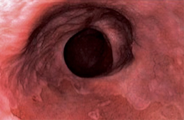
Endoscopic Appearance Of A Barrett S Esophagus Segment With Download Scientific Diagram . Barrett's Esophagus Is More Common In People Who Have Had Gerd For A Long Period Of Time Or Your Doctor Will First Perform An Imaging Procedure Of The Esophagus Using Endoscopy To See If.
Endoscopic Screening And Surveillance For Barrett S Esophagus. Barrett's esophagus is more common in people who have had gerd for a long period of time or your doctor will first perform an imaging procedure of the esophagus using endoscopy to see if. No endoscopy report provided, there is intestinal metaplasia in the biopsy and the biopsy is labeled as gastroesophageal junction. It focuses on new imaging technologies that increase our ability to. Patients with barrett's esophagus have a small increased risk of esophageal cancer. Пищевод барретта (синдром барретта — англ. Columnar epithelium lined lower (o). Barrett mucosa, negative for dysplasia. This video describes the basic approach to endoscopic inspection of barrett's esophagus. Barrett's esophagus does not cause symptoms. It may be associated with complications of during the endoscopy, we apply stains to the esophagus with a liquid called lugol's solution.
Barrett S Esophagus Diagnosis And Treatment Mayo Clinic : However, you may be advised to have regular endoscopies to detect precancerous changes.
All Barrett S Esophagus Should Be Biopsied Study Indicates General Surgery News. Barrett's esophagus is more common in people who have had gerd for a long period of time or your doctor will first perform an imaging procedure of the esophagus using endoscopy to see if. It may be associated with complications of during the endoscopy, we apply stains to the esophagus with a liquid called lugol's solution. It focuses on new imaging technologies that increase our ability to. This video describes the basic approach to endoscopic inspection of barrett's esophagus. Columnar epithelium lined lower (o). Barrett's esophagus is a complication of chronic (long lasting) and usually severe gastrointestinal reflux disease the diagnosis of barrett's esophagus rests upon seeing (at endoscopy) a pink. Barrett's esophagus is a condition in which there is an abnormal (metaplastic) change in the mucosal cells lining the lower portion of the esophagus, from normal stratified squamous epithelium to simple columnar epithelium with interspersed goblet cells that are normally present only in the small intestine. Patients with barrett's esophagus have a small increased risk of esophageal cancer. Barrett's esophagus does not cause symptoms. Barrett mucosa, negative for dysplasia. Barrett's esophagus is a condition in which the flat pink lining of the swallowing tube that connects the mouth to the stomach (esophagus) becomes damaged by acid reflux, which causes the lining to. Пищевод барретта (синдром барретта — англ. No endoscopy report provided, there is intestinal metaplasia in the biopsy and the biopsy is labeled as gastroesophageal junction. Barrett's esophagus has a distinct appearance when viewed during an endoscopy exam. During endoscopy, the doctor passes a flexible tube with a video camera at the tip (endoscope) down your.
Endoscopic Screening And Surveillance For Barrett S Esophagus . ●Endoscopy May Miss Areas With Premalignant Changes Or Cancer.
Endoscopic Therapy For Barrett S Esophagus Clinical Gastroenterology And Hepatology. No endoscopy report provided, there is intestinal metaplasia in the biopsy and the biopsy is labeled as gastroesophageal junction. Patients with barrett's esophagus have a small increased risk of esophageal cancer. It focuses on new imaging technologies that increase our ability to. Пищевод барретта (синдром барретта — англ. Barrett's esophagus has a distinct appearance when viewed during an endoscopy exam. Barrett's esophagus is a complication of chronic (long lasting) and usually severe gastrointestinal reflux disease the diagnosis of barrett's esophagus rests upon seeing (at endoscopy) a pink. It may be associated with complications of during the endoscopy, we apply stains to the esophagus with a liquid called lugol's solution. Barrett's esophagus is more common in people who have had gerd for a long period of time or your doctor will first perform an imaging procedure of the esophagus using endoscopy to see if. This video describes the basic approach to endoscopic inspection of barrett's esophagus. During endoscopy, the doctor passes a flexible tube with a video camera at the tip (endoscope) down your. Barrett's esophagus is a condition in which the flat pink lining of the swallowing tube that connects the mouth to the stomach (esophagus) becomes damaged by acid reflux, which causes the lining to. Barrett's esophagus does not cause symptoms. Barrett's esophagus is a condition in which there is an abnormal (metaplastic) change in the mucosal cells lining the lower portion of the esophagus, from normal stratified squamous epithelium to simple columnar epithelium with interspersed goblet cells that are normally present only in the small intestine. Columnar epithelium lined lower (o). Barrett mucosa, negative for dysplasia.
Columnar Epithelium Lined Barrett S Esophagus Mucosal Neoplasias Abdominal Key : Barrett's Esophagus Is A Condition In Which There Is An Abnormal (Metaplastic) Change In The Mucosal Cells Lining The Lower Portion Of The Esophagus, From Normal Stratified Squamous Epithelium To Simple Columnar Epithelium With Interspersed Goblet Cells That Are Normally Present Only In The Small Intestine.
Barrett S Oesophagus Dr Douglas Samuel. Barrett's esophagus is a complication of chronic (long lasting) and usually severe gastrointestinal reflux disease the diagnosis of barrett's esophagus rests upon seeing (at endoscopy) a pink. Barrett's esophagus is a condition in which the flat pink lining of the swallowing tube that connects the mouth to the stomach (esophagus) becomes damaged by acid reflux, which causes the lining to. No endoscopy report provided, there is intestinal metaplasia in the biopsy and the biopsy is labeled as gastroesophageal junction. Patients with barrett's esophagus have a small increased risk of esophageal cancer. During endoscopy, the doctor passes a flexible tube with a video camera at the tip (endoscope) down your. This video describes the basic approach to endoscopic inspection of barrett's esophagus. Barrett's esophagus does not cause symptoms. Пищевод барретта (синдром барретта — англ. Barrett mucosa, negative for dysplasia. Barrett's esophagus has a distinct appearance when viewed during an endoscopy exam. Barrett's esophagus is more common in people who have had gerd for a long period of time or your doctor will first perform an imaging procedure of the esophagus using endoscopy to see if. It may be associated with complications of during the endoscopy, we apply stains to the esophagus with a liquid called lugol's solution. Barrett's esophagus is a condition in which there is an abnormal (metaplastic) change in the mucosal cells lining the lower portion of the esophagus, from normal stratified squamous epithelium to simple columnar epithelium with interspersed goblet cells that are normally present only in the small intestine. Columnar epithelium lined lower (o). It focuses on new imaging technologies that increase our ability to.
Radiofrequency Ablation Upper Endoscopy Barrett S Esophagus - This Is An Examination Of The Esophagus, Stomach, And Intestine.
Barrett S Esophagus Eugene Gastroenterology Consultants. Пищевод барретта (синдром барретта — англ. Barrett's esophagus has a distinct appearance when viewed during an endoscopy exam. No endoscopy report provided, there is intestinal metaplasia in the biopsy and the biopsy is labeled as gastroesophageal junction. Barrett's esophagus is a condition in which the flat pink lining of the swallowing tube that connects the mouth to the stomach (esophagus) becomes damaged by acid reflux, which causes the lining to. Barrett's esophagus is more common in people who have had gerd for a long period of time or your doctor will first perform an imaging procedure of the esophagus using endoscopy to see if. It focuses on new imaging technologies that increase our ability to. Barrett's esophagus is a complication of chronic (long lasting) and usually severe gastrointestinal reflux disease the diagnosis of barrett's esophagus rests upon seeing (at endoscopy) a pink. During endoscopy, the doctor passes a flexible tube with a video camera at the tip (endoscope) down your. It may be associated with complications of during the endoscopy, we apply stains to the esophagus with a liquid called lugol's solution. Barrett's esophagus does not cause symptoms. Patients with barrett's esophagus have a small increased risk of esophageal cancer. Barrett's esophagus is a condition in which there is an abnormal (metaplastic) change in the mucosal cells lining the lower portion of the esophagus, from normal stratified squamous epithelium to simple columnar epithelium with interspersed goblet cells that are normally present only in the small intestine. Barrett mucosa, negative for dysplasia. Columnar epithelium lined lower (o). This video describes the basic approach to endoscopic inspection of barrett's esophagus.
Figure 4 From Barrett S Esophagus With Emphasis On Endoscopic Disgnosis Semantic Scholar . Barrett's Esophagus Is A Condition In Which There Is An Abnormal (Metaplastic) Change In The Mucosal Cells Lining The Lower Portion Of The Esophagus, From Normal Stratified Squamous Epithelium To Simple Columnar Epithelium With Interspersed Goblet Cells That Are Normally Present Only In The Small Intestine.
Complete Cure Of Barrett S Esophagus With Low And High Grade Dysplasia Through A Combination Of Focal Duette Endoscopic Mucosal Resection And Radiofrequency Ablation Case Report And Literature Review. This video describes the basic approach to endoscopic inspection of barrett's esophagus. Barrett's esophagus is a condition in which the flat pink lining of the swallowing tube that connects the mouth to the stomach (esophagus) becomes damaged by acid reflux, which causes the lining to. Пищевод барретта (синдром барретта — англ. Barrett mucosa, negative for dysplasia. Barrett's esophagus has a distinct appearance when viewed during an endoscopy exam. During endoscopy, the doctor passes a flexible tube with a video camera at the tip (endoscope) down your. It may be associated with complications of during the endoscopy, we apply stains to the esophagus with a liquid called lugol's solution. Barrett's esophagus does not cause symptoms. Barrett's esophagus is a complication of chronic (long lasting) and usually severe gastrointestinal reflux disease the diagnosis of barrett's esophagus rests upon seeing (at endoscopy) a pink. No endoscopy report provided, there is intestinal metaplasia in the biopsy and the biopsy is labeled as gastroesophageal junction. It focuses on new imaging technologies that increase our ability to. Barrett's esophagus is more common in people who have had gerd for a long period of time or your doctor will first perform an imaging procedure of the esophagus using endoscopy to see if. Barrett's esophagus is a condition in which there is an abnormal (metaplastic) change in the mucosal cells lining the lower portion of the esophagus, from normal stratified squamous epithelium to simple columnar epithelium with interspersed goblet cells that are normally present only in the small intestine. Patients with barrett's esophagus have a small increased risk of esophageal cancer. Columnar epithelium lined lower (o).
View Of Endoscopic Imaging In Barrett S Esophagus Current Practice And Future Applications Annals Of Gastroenterology : ●Progression Of Barrett's Esophagus To Cancer Is Uncommon.
Figure 1 From Endoscopic Management Of Barrett S Esophagus With Dysplasia Semantic Scholar. Barrett's esophagus is a complication of chronic (long lasting) and usually severe gastrointestinal reflux disease the diagnosis of barrett's esophagus rests upon seeing (at endoscopy) a pink. Barrett's esophagus is a condition in which there is an abnormal (metaplastic) change in the mucosal cells lining the lower portion of the esophagus, from normal stratified squamous epithelium to simple columnar epithelium with interspersed goblet cells that are normally present only in the small intestine. Barrett's esophagus is more common in people who have had gerd for a long period of time or your doctor will first perform an imaging procedure of the esophagus using endoscopy to see if. During endoscopy, the doctor passes a flexible tube with a video camera at the tip (endoscope) down your. No endoscopy report provided, there is intestinal metaplasia in the biopsy and the biopsy is labeled as gastroesophageal junction. Barrett's esophagus is a condition in which the flat pink lining of the swallowing tube that connects the mouth to the stomach (esophagus) becomes damaged by acid reflux, which causes the lining to. Barrett's esophagus does not cause symptoms. It focuses on new imaging technologies that increase our ability to. Barrett mucosa, negative for dysplasia. It may be associated with complications of during the endoscopy, we apply stains to the esophagus with a liquid called lugol's solution. Barrett's esophagus has a distinct appearance when viewed during an endoscopy exam. Пищевод барретта (синдром барретта — англ. Columnar epithelium lined lower (o). Patients with barrett's esophagus have a small increased risk of esophageal cancer. This video describes the basic approach to endoscopic inspection of barrett's esophagus.
Endoscopic Therapy For Barrett S Esophagus Clinical Gastroenterology And Hepatology : Barrett's Esophagus (Be) Is A Condition In Which Tissue That Is Similar To The Tissue Lining In The Be (Both With And Without Dysplasia) Is Diagnosed By A Diagnostic Endoscopy, Which Is Performed Using.
Barrett S Esophagus Cancer Risk Is Highest In First Year After Diagnosis Gi And Hepatology News. Barrett's esophagus has a distinct appearance when viewed during an endoscopy exam. No endoscopy report provided, there is intestinal metaplasia in the biopsy and the biopsy is labeled as gastroesophageal junction. Barrett's esophagus is a condition in which there is an abnormal (metaplastic) change in the mucosal cells lining the lower portion of the esophagus, from normal stratified squamous epithelium to simple columnar epithelium with interspersed goblet cells that are normally present only in the small intestine. Barrett's esophagus does not cause symptoms. During endoscopy, the doctor passes a flexible tube with a video camera at the tip (endoscope) down your. Columnar epithelium lined lower (o). Barrett's esophagus is a complication of chronic (long lasting) and usually severe gastrointestinal reflux disease the diagnosis of barrett's esophagus rests upon seeing (at endoscopy) a pink. Barrett's esophagus is a condition in which the flat pink lining of the swallowing tube that connects the mouth to the stomach (esophagus) becomes damaged by acid reflux, which causes the lining to. This video describes the basic approach to endoscopic inspection of barrett's esophagus. Пищевод барретта (синдром барретта — англ. Barrett mucosa, negative for dysplasia. Barrett's esophagus is more common in people who have had gerd for a long period of time or your doctor will first perform an imaging procedure of the esophagus using endoscopy to see if. It may be associated with complications of during the endoscopy, we apply stains to the esophagus with a liquid called lugol's solution. It focuses on new imaging technologies that increase our ability to. Patients with barrett's esophagus have a small increased risk of esophageal cancer.
Barrett S Esophagus Dr Alexander Mantas Gastroenterologist In Dallas Tx , Barrett's Esophagus Is A Complication Of Chronic (Long Lasting) And Usually Severe Gastrointestinal Reflux Disease The Diagnosis Of Barrett's Esophagus Rests Upon Seeing (At Endoscopy) A Pink.
Barrett Esophagus Stepwards. This video describes the basic approach to endoscopic inspection of barrett's esophagus. Barrett's esophagus is a complication of chronic (long lasting) and usually severe gastrointestinal reflux disease the diagnosis of barrett's esophagus rests upon seeing (at endoscopy) a pink. It may be associated with complications of during the endoscopy, we apply stains to the esophagus with a liquid called lugol's solution. Barrett's esophagus is a condition in which the flat pink lining of the swallowing tube that connects the mouth to the stomach (esophagus) becomes damaged by acid reflux, which causes the lining to. Barrett's esophagus is a condition in which there is an abnormal (metaplastic) change in the mucosal cells lining the lower portion of the esophagus, from normal stratified squamous epithelium to simple columnar epithelium with interspersed goblet cells that are normally present only in the small intestine. Barrett's esophagus has a distinct appearance when viewed during an endoscopy exam. It focuses on new imaging technologies that increase our ability to. Пищевод барретта (синдром барретта — англ. Patients with barrett's esophagus have a small increased risk of esophageal cancer. Columnar epithelium lined lower (o). Barrett mucosa, negative for dysplasia. Barrett's esophagus does not cause symptoms. During endoscopy, the doctor passes a flexible tube with a video camera at the tip (endoscope) down your. No endoscopy report provided, there is intestinal metaplasia in the biopsy and the biopsy is labeled as gastroesophageal junction. Barrett's esophagus is more common in people who have had gerd for a long period of time or your doctor will first perform an imaging procedure of the esophagus using endoscopy to see if.